Stay Up to Date
Submit your email address to receive the latest industry and Aerospace America news.

Additive manufacturing, also called 3-D printing, can’t revolutionize aviation until engineers can print parts with repeatable quality and specifications, and consistent mechanical properties. One hurdle has been a near inability to see how irregularities form within particular metals. Researchers at Argonne National Laboratory outside Chicago are addressing that challenge by watching 3-D manufacturing with powerful X-rays. Argonne’s Aaron Greco and Tao Sun tell the story.
A challenging problem in aerospace engineering is: How can we build metal parts with improved functionalities and performance without increasing their weight? Additive manufacturing or AM for short, is one potential solution. AM or 3-D printing refers to a suite of techniques for building three-dimensional objects by adding materials layer by layer based on a digital design. AM structures can be topologically optimized to reduce the weight of parts in planes, thereby saving fuel and decreasing CO2 emissions. AM also largely eliminates tooling constraints, and gives engineers the freedom to design and build parts with complex geometries and improved performance. Other advantages of AM include short time to market, a short supply chain, and on-site manufacturing of spares.
As promising as AM is, there are still many fundamental problems we need to solve before these techniques can reach their full potential of revolutionizing the way we build parts for aircraft. A key challenge is that only a few metals are currently considered suitable for use in AM after years-long efforts on process optimization. For others, we do not understand yet how to reliably produce parts without cracks, porosities, and other microstructure defects. At Argonne National Laboratory outside Chicago, we (a team with backgrounds in physics, materials science, computer science and engineering) began tackling this problem in 2016 by additively manufacturing metal parts in the path of a high powered X-ray to observe how various defects form.
To understand why AM metal parts are susceptible to defects, it’s helpful to consider the process. An electron or laser beam melts a feedstock, most typically in the form of a powder. In laser powder bed fusion, a thin bed of metal powder is mechanically spread on top of the previously fused layer. The laser melts the powder and the previously printed layer. Several problems can occur before the material cools and fuses. The extremely high and non-uniform thermal gradient inside the sample leads to a high surface tension difference, making conditions favorable for Marangoni flow, in which a gradient or difference in surface tension causes liquid material to move. In the case of laser fusion, liquid flows from the center of the laser-heated zone (lower surface tension) to the surrounding regions (higher surface tension), and so does the heat. As a result, the melt depth increases, and the powders in the close vicinity are pulled into the melt pool, and high-speed surface liquid metal may break away from the melt pool and spatter off. Meanwhile, the heat also vaporizes some of the metal, creating a small cavity, called a vapor depression zone. The vapor carries away some amount of powders and molten metal from the powder bed. The speed of the ejected powder can reach tens of meters per second. Once the laser leaves the area, the temperature decreases and the sample solidifies rapidly, sometimes accompanied by phase transformation (i.e., change in the atomistic structures of metals) and/or precipitation of a secondary phase (i.e., a certain part of the material contains different crystal structure with the surrounding matrix).
The entire melting and solidification process typically lasts only a few milliseconds in powder bed AM, so capturing these transient phenomena with high spatial and temporal resolution has been very challenging. Scientists have achieved some success in the past by illuminating the process with visible light and taking high-speed images. Powder flow, ejection, and melt pool dynamics near the sample surface can all be observed using this technique. However, almost all metals are opaque, so we cannot use visible light imaging to watch what happens below the surface and inside the sample during printing, and this is where most of the microstructure evolution occurs.
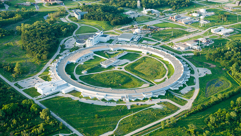
Argonne is the home of the Advanced Photon Source, or APS. The APS is a synchrotron facility, which provides ultra-bright, high-energy storage ring-generated X-ray beams for research in almost all scientific disciplines. In the storage ring, electrons with energy of 7 billion electron volts are traveling at greater than 99.999999 percent of the speed of light. When these moving electrons change direction, they emit energy at X-ray wavelength. Scientists have known about the unique ability of X-rays to penetrate solid matter ever since Wilhelm Röntgen first discovered X-rays more than 100 years ago. The APS X-rays are about a billion times brighter than those generated by a machine in a hospital’s radiology department. For synchrotron facilities, the larger the circumference of the storage ring, the higher the energy of electrons can reach, and the more flux the high-energy X-rays can contain. The APS is one of four high-energy synchrotron facilities in the world. The others are in Japan, France, and Germany.
In fiscal 2016, Argonne provided internal research and development funds to support our project to study metal AM with X-ray imaging. These development funds are intended for high-technical risk, yet potentially high-payoff research and development projects like ours. Frankly, even though we knew how to build the laser AM apparatus and conduct the X-ray experiment, we had very limited ideas about the kinds of phenomena we would be able to see. Therefore, at the beginning of this project, we only hoped to get some images showing the dynamics of powder melting while we heated it with a short-pulse laser. We started the project by building our own laser AM system. With the initial funding, we purchased a high-power laser that was delivered in May 2016, followed in July by a custom-built sample chamber that would be placed in the X-ray beamline. We then faced a challenge — we didn’t have enough funding left to buy the laser optics. In the end, IPG Photonics Corp., from which we purchased our laser source, was kind enough to lend us a laser head, so our project could proceed.
In August 2016, we performed the first high-speed X-ray imaging experiment at the beamline of the APS. We had six days of beam time in August, the last month of the final APS cycle for the fiscal year. The pressure was pretty high at that point. If we couldn’t get results from our experiment, we would end our first-year project almost empty handed, because the APS shuts down every September. We wouldn’t have a chance to try again until it reopened one and a half months later, at the start of fiscal 2017. We spent two and a half days assembling the laser system, integrating it into the beamline, and certifying it with Argonne laser safety officials. On the afternoon of Aug. 20, right after lunch, we opened the X-ray shutter and performed our first high-speed imaging experiment on laser heating process. We chose a titanium alloy plate to start with. The result didn’t disappoint us at all. We could clearly see the melt pool developing inside the sample as the laser heated it. After a few adjustments of the laser beam size, we began to observe additional phenomena, including the dynamics of vapor depression zones, Marangoni flows of molten materials, and rapid solidification. On Aug. 21, we started to collect high-quality X-ray images of the powder bed samples. On Aug. 22, we got greedier. We set up a diffraction detector and conducted simultaneous imaging and diffraction to probe the phase transformation process in titanium alloy. By the end of our beam time, we had collected nearly 90 gigabytes of data from 150 samples. We summarized these data and published them in Scientific Reports.
In fiscal 2017, we received extra funding from Argonne and bought a laser scanner that functions just like those in commercial powder bed fusion machines. We began to collaborate with a team led by Anthony Rollett, a professor at Carnegie Mellon University, and one led by Lianyi Chen, a professor at Missouri University of Science and Technology. Together, we designed and conducted X-ray experiments to investigate fundamental problems in laser powder bed fusion associated with porosity and crack generation, melt pool flow, solidification and powder ejection. Our collaborative team is currently drafting several papers summarizing our observations and the understanding we gained on these topics.
Along with the experiments, we have been developing multiple image analysis approaches to extract as much microstructure information from our X-ray data as possible. With the quantitative data about the dynamic material microstructure evolution during AM processes, we can help build highly accurate numerical models to optimize the manufacturing of parts with different geometries and dimensions. Metal AM has developed rapidly in recent years, thanks to substantial investments in the technology from both public and private groups worldwide. Gaining precise control of microstructures and the properties of additively manufactured products remains challenging, which is why at this writing the pool of materials for AM remains very small. We believe our research delivers new information about the physics underpinning metal AM process, facilitates the development of new alloys for AM, and accelerates the application of AM in the aerospace industry and many other fields.
Related Posts
Stay Up to Date
Submit your email address to receive the latest industry and Aerospace America news.